Hemorrhage, sepsis, medication effects, dysrhythmia, or pump failure can reduce driving pressure.
CHAPTER5
Placental and Fetal Physiology
Learning objectives
After completing this chapter, you should be able to:
- Describe the separate maternal and fetal circulations within the placenta.
- Explain how maternal cardiac output, perfusion pressure, oxygen content, uterine blood flow, and placental function determine fetal oxygen delivery.
- Trace fetal blood through the umbilical vein, ductus venosus, foramen ovale, ductus arteriosus, and umbilical arteries.
- Explain why the fetal ventricles function largely in parallel and why pulmonary vascular resistance is high before birth.
- Describe how fetal hemoglobin, blood-flow streaming, and redistribution support oxygen delivery to vital organs.
- Recognize the progression from fetal compensation to decompensation during maternal or placental compromise.
- Apply maternal positioning, oxygenation, perfusion, monitoring, communication, and diversion principles to fetal deterioration during transport.
Opening transport scenario
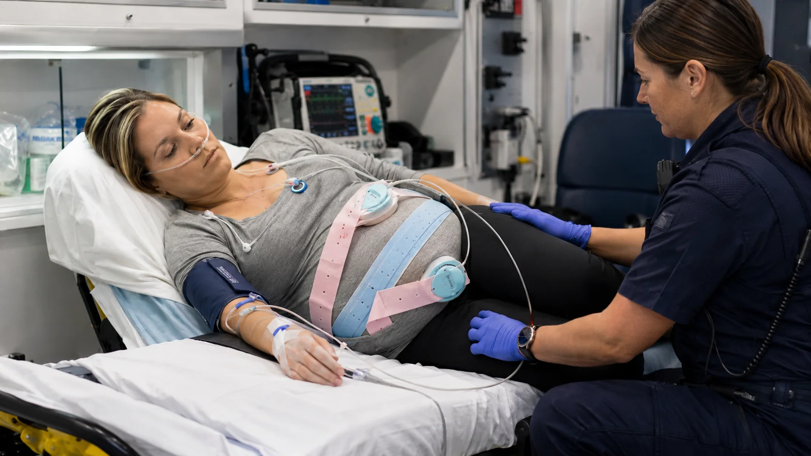
A 31-year-old patient at 29 weeks’ gestation is being transferred for severe fetal growth restriction and abnormal umbilical artery Doppler findings. At departure, maternal vital signs are stable, fetal heart rate is 145/min with moderate variability, and the patient is positioned with a left lateral tilt. Twenty minutes into transport, vomiting and anxiety are followed by maternal hypoxemia and a blood-pressure decline. The fetal tracing develops recurrent late decelerations and then prolonged bradycardia.
The fetal monitor is displaying the downstream effect of a larger system failure. Fetal oxygen delivery depends on the maternal lungs, maternal hemoglobin, maternal cardiac output, uterine perfusion, placental exchange, umbilical blood flow, fetal hemoglobin, and fetal cardiovascular distribution. A problem at any point can reduce oxygen delivery. The transport team must stabilize the mother, assess the fetal response, and determine whether the original destination remains appropriate.
The maternal–placental–fetal system
Five links must remain intact
1Maternal oxygen contentVentilation, oxygen saturation, hemoglobin
2Maternal cardiac outputPreload, heart rate, stroke volume
3Uterine blood flowPerfusion pressure, position, vascular tone
4Placental exchangeSurface area, diffusion distance, vascular resistance
5Fetal distributionUmbilical flow, fetal hemoglobin, cardiac shunts
1. The placenta: a temporary multiorgan support system
The placenta is not simply an attachment between mother and fetus. It functions as the fetal lungs, gastrointestinal interface, kidneys, endocrine organ, and immunologic boundary. It transfers oxygen and nutrients to the fetus, removes carbon dioxide and metabolic waste, produces hormones that alter maternal physiology, and helps regulate fetal growth.
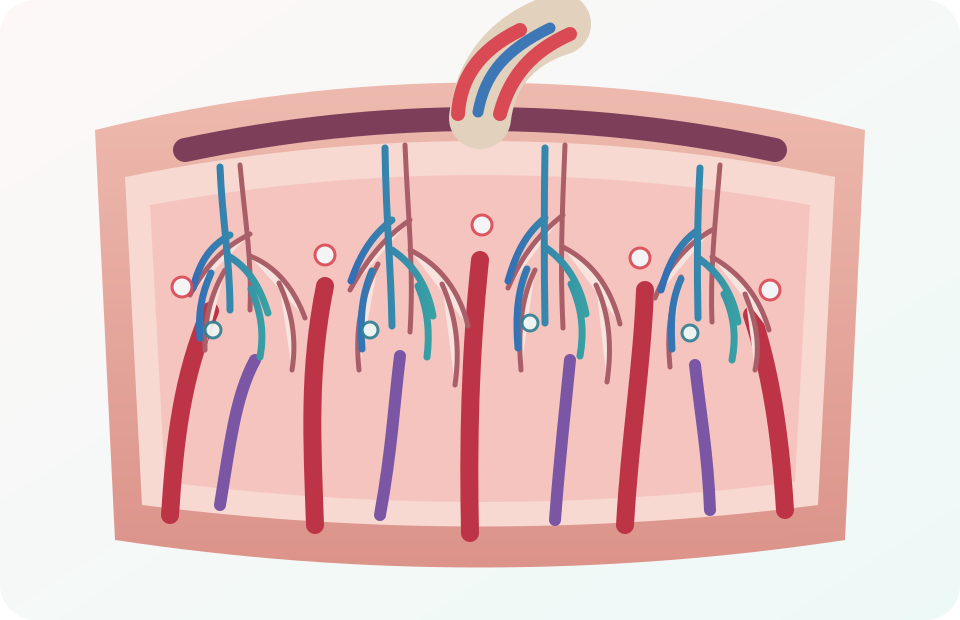
Two circulations, one exchange surface
Maternal side
Spiral arteries deliver maternal blood into the intervillous space. Uterine veins drain it back to the maternal circulation.
Fetal side
Umbilical arteries deliver fetal blood to villous capillaries. The umbilical vein returns the most oxygenated fetal blood.
1.1 Chorionic villi and intervillous blood
Chorionic villi create an extensive branching exchange surface. Fetal capillaries run within the villi while maternal blood surrounds them in the intervillous space. Efficient exchange depends on adequate flow on both sides, a large functional surface area, a short diffusion distance, and favorable concentration gradients.
1.2 Placental endocrine and metabolic roles
The placenta produces hormones and signaling molecules that help maintain pregnancy and adapt maternal metabolism. It also transports glucose, amino acids, fatty acids, electrolytes, and micronutrients using diffusion, facilitated diffusion, and active transport. Placental transport is selective, but it is not a perfect barrier; many medications and toxins can cross.
2. Uteroplacental circulation
Normal placentation transforms the maternal spiral arteries into high-capacitance, low-resistance vessels. Near term, the uterus and placenta receive a substantial fraction of maternal cardiac output. Uteroplacental blood flow has limited autoregulatory capacity and depends strongly on maternal perfusion pressure, maternal cardiac output, uterine vascular resistance, and maternal position.
1Maternal cardiac outputFlow generated by the maternal heart
→2Perfusion pressurePressure available to drive uterine flow
→3Spiral artery flowLow-resistance delivery to the intervillous space
→4Placental exchangeGas and substrate transfer
→5Fetal oxygen deliveryUmbilical flow and fetal distribution
2.1 Factors that reduce uterine blood flow
Supine positioning can reduce venous return, cardiac output, and uterine perfusion.
Even preserved flow may deliver inadequate oxygen content.
Catecholamine surge, stimulant use, severe pain, or some medications can reduce uterine flow.
Frequent or prolonged contractions shorten the time available for placental reperfusion.
Abruption, infarction, abnormal implantation, or impaired vascular development reduce functional exchange.
2.2 Contractions and placental reperfusion
During a contraction, increased uterine pressure compresses maternal vessels and transiently reduces intervillous perfusion. A healthy fetus normally tolerates this because oxygen is stored in fetal blood and tissues, and flow recovers between contractions. Tachysystole, prolonged contractions, maternal hypotension, or placental disease can prevent adequate recovery and produce recurrent fetal heart-rate abnormalities.
Why can left lateral positioning improve fetal status even when no fetal medication is given?
Answer: It can relieve vena-caval compression, improve maternal preload and cardiac output, increase uterine perfusion, and therefore improve placental oxygen transfer.
3. Umbilical cord physiology
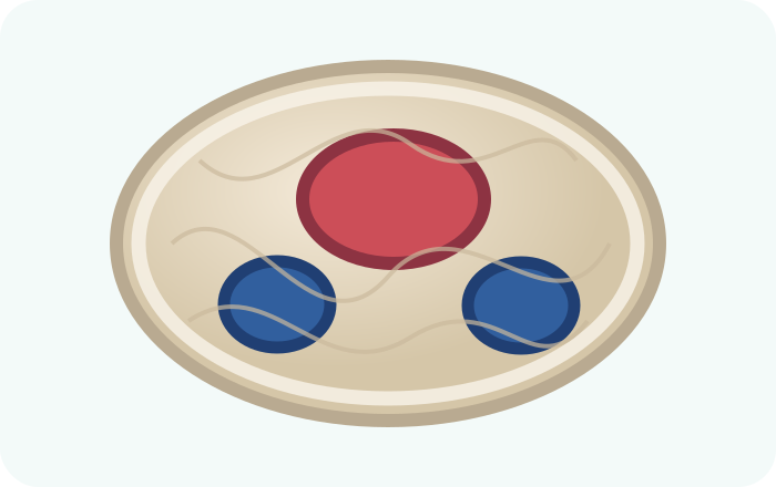
Vessel direction, not oxygen content
Umbilical vein
Returns oxygenated, nutrient-rich blood from placenta to fetus.
Two umbilical arteries
Carry lower-oxygen blood and waste from fetus to placenta.
Wharton jelly
Protective connective tissue that helps resist compression and kinking.
3.1 Cord compression
Cord compression can reduce blood flow through the thin-walled umbilical vein and, with greater compression, the arteries. Intermittent compression classically produces variable decelerations. Persistent compression, prolapse, knotting, entanglement, or oligohydramnios can produce sustained fetal compromise.
3.2 Transport implications
- Avoid unnecessary pressure over the gravid abdomen.
- Secure the patient without compressing the uterus or dislodging fetal-monitor transducers.
- Recognize abrupt fetal heart-rate changes after patient movement, stretcher loading, or rupture of membranes.
- When cord prolapse is suspected, prioritize pressure relief, maternal positioning, immediate obstetric communication, and rapid delivery capability according to protocol.
4. Fetal circulation
Fetal circulation is arranged around placental—not pulmonary—gas exchange. The ventricles contribute to a combined cardiac output with substantial parallel flow. Three shunts direct blood around organs that are not yet functioning in their postnatal roles.
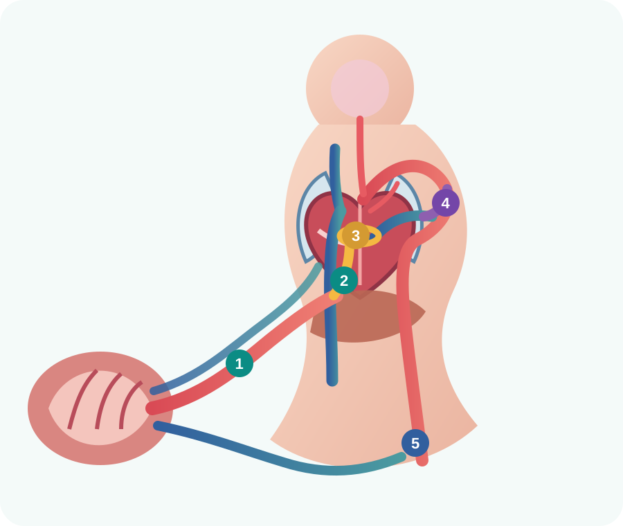
1Umbilical vein
Brings the highest-oxygen fetal blood from the placenta.
2Ductus venosus
Directs a portion of umbilical venous blood toward the inferior vena cava, bypassing much of the liver.
3Foramen ovale
Streams relatively oxygen-rich blood from the right atrium to the left atrium and then toward the brain and coronary circulation.
4Ductus arteriosus
Diverts most pulmonary artery blood into the descending aorta because fetal pulmonary vascular resistance is high.
5Umbilical arteries
Return lower-oxygen blood to the placenta for gas and waste exchange.
4.1 Preferential streaming
Oxygen-rich inferior vena-caval blood is preferentially directed across the foramen ovale toward the left atrium, left ventricle, ascending aorta, coronary arteries, and brain. Blood returning through the superior vena cava is more likely to enter the right ventricle and pulmonary artery, then pass through the ductus arteriosus to the descending aorta.
4.2 Why pulmonary flow is low before birth
The fetal lungs are fluid-filled and unexpanded, and alveolar oxygen tension is low. Pulmonary vascular resistance remains high, so only a minority of right-ventricular output flows through the lungs. At birth, lung expansion and rising oxygen tension reduce pulmonary vascular resistance, initiating the transition covered in Chapter 6.
Ductus venosus
Placenta → umbilical vein → IVC pathway that partially bypasses liver.
Foramen ovale
Right atrium → left atrium; prioritizes upper-body oxygen delivery.
Ductus arteriosus
Pulmonary artery → descending aorta; bypasses high-resistance lungs.
Which fetal shunt directly routes blood from the pulmonary artery into the aorta?
Answer: The ductus arteriosus.
5. Fetal oxygen transport
Fetal arterial oxygen tension is lower than adult arterial oxygen tension, yet fetal tissues receive adequate oxygen through several adaptations: fetal hemoglobin has higher oxygen affinity, fetal hemoglobin concentration is relatively high, cardiac output per kilogram is high, and blood flow is preferentially distributed toward vital organs.
Fetal hemoglobin binds oxygen effectively at lower partial pressures.
More hemoglobin supports arterial oxygen content despite lower PaO₂.
Flow helps compensate for lower oxygen tension.
Relatively oxygen-rich blood is directed toward brain and myocardium.
5.1 Placental gas transfer
Oxygen diffuses from maternal blood to fetal blood, while carbon dioxide moves in the opposite direction. Maternal unloading and fetal loading are supported by differences in hemoglobin affinity and the exchange of carbon dioxide and hydrogen ions across the placenta. Efficient transfer still requires adequate flow and functional placental surface area.
5.2 Oxygen delivery equation applied to the fetus
Fetal oxygen delivery=Umbilical blood flow×Fetal arterial oxygen content
A problem can therefore result from reduced flow, reduced oxygen content, or both. Maternal hypoxemia reduces the oxygen gradient. Maternal anemia reduces available oxygen content. Maternal hypotension reduces uterine flow. Placental disease increases resistance or reduces exchange area. Cord compression reduces umbilical flow.
6. Fetal response to hypoxemia
The fetus has compensatory mechanisms but limited reserve. The clinical trajectory depends on severity, duration, gestational age, placental reserve, and the ability to restore maternal and placental conditions.
1Compensation
→Catecholamine release, redistribution toward brain/heart/adrenals, altered movement, and fetal heart-rate changes.
2Threatened reserve
→Persistent decelerations, reduced variability, increasing anaerobic metabolism, and accumulating lactate.
3Decompensation
Myocardial depression, sustained bradycardia, severe acidosis, hypotension, and loss of protective redistribution.
6.1 Brain-sparing is not reassurance
Redistribution toward the brain may temporarily preserve essential organs in chronic placental insufficiency. It is evidence of adaptation to stress—not proof that the underlying condition is safe. Severe or prolonged compromise eventually overwhelms the response.
6.2 Fetal heart-rate changes as physiology
Fetal heart-rate patterns reflect autonomic control, oxygenation, acid-base status, medications, sleep cycles, gestational age, and mechanical influences such as head or cord compression. A tracing should be interpreted as a trend in the context of maternal physiology and contractions, not as an isolated number.
7. Placental resistance and Doppler concepts
Umbilical artery Doppler evaluates resistance within the fetoplacental circulation. Normal placental development produces progressively lower resistance and forward end-diastolic flow. Decreased, absent, or reversed end-diastolic flow indicates increasing placental resistance and is associated with placental insufficiency and fetal growth restriction.
Forward diastolic flow
Expected when placental resistance is adequately low.
Absent end-diastolic flow
Severe placental resistance and increased risk of deterioration.
Reversed end-diastolic flow
Advanced compromise requiring heightened surveillance and delivery planning.
Transport clinicians may receive Doppler results from the sending facility rather than perform the study themselves. These results should influence urgency, destination capability, monitoring, corticosteroid or magnesium planning when ordered, and the threshold for escalation.
8. Maternal events and predicted fetal effects
| Maternal or placental event | Primary physiologic effect | Possible fetal consequence |
|---|---|---|
| Maternal hypoxemia | Lower oxygen available for placental diffusion | Tachycardia, decelerations, bradycardia, acidosis |
| Maternal hypotension | Reduced uterine perfusion pressure | Late decelerations, reduced variability, prolonged bradycardia |
| Severe anemia or hemorrhage | Reduced maternal oxygen content and circulating volume | Fetal hypoxemia before dramatic maternal hypotension |
| Supine aortocaval compression | Reduced venous return and cardiac output | Rapid fetal deterioration that may improve with position |
| Uterine tachysystole | Inadequate reperfusion between contractions | Recurrent late or prolonged decelerations |
| Placental abruption | Loss of functional exchange area plus maternal hemorrhage | Acute hypoxia, bradycardia, fetal death |
| Maternal fever or sepsis | Higher metabolic demand, vasodilation, possible hypoperfusion | Fetal tachycardia and progressive compromise |
| Cord compression or prolapse | Reduced umbilical venous and arterial flow | Variable decelerations or sustained bradycardia |
9. Transport assessment and interventions
Optimize maternal oxygenation
Assess airway, work of breathing, SpO₂, ventilation, hemoglobin context, and oxygen reserve.
Optimize maternal perfusion
Correct position, hemorrhage, hypotension, dysrhythmia, or shock according to cause.
Protect uterine flow
Use left tilt when appropriate, minimize unnecessary vasoconstriction, and reassess during movement.
Trend fetal response
Verify fetal versus maternal heart rate, assess variability and decelerations when monitoring is available, and document changes.
Communicate early
Report maternal changes, fetal trajectory, interventions, response, travel time, and alternative destinations.
Prepare for failure
Plan for urgent delivery, neonatal needs, diversion, airway deterioration, hemorrhage, and equipment limitations.

9.1 Verify the signal
External fetal monitors can display artifact during movement. Compare the fetal rate with the maternal pulse or ECG, assess transducer placement, listen with Doppler when appropriate, and treat a persistent concerning rate as real until verified otherwise. Do not allow artifact concerns to delay escalation when the clinical picture is deteriorating.
9.2 Treat the maternal mechanism
There is no universal “fetal resuscitation drug” for transport. Correct the maternal cause: relieve vena-caval compression, restore oxygenation and ventilation, control hemorrhage, support perfusion, treat seizure or sepsis, stop a causative medication when ordered, reduce excessive uterine activity according to protocol, and expedite obstetric capability.
10. Evolving transport case
Phase 1: Known placental disease
The patient has severe fetal growth restriction with absent end-diastolic flow. This indicates high placental resistance and limited reserve. The team confirms continuous fetal monitoring, emergency delivery capability at the destination, antenatal medication history, neonatal readiness, and the closest alternate obstetric facility.
Phase 2: Maternal deterioration
Vomiting is followed by SpO₂ 88%, blood pressure 84/50 mm Hg, and fetal late decelerations. The team positions the patient laterally, administers oxygen, assesses ventilation, verifies lines and medications, evaluates for hemorrhage or medication effect, and communicates the change.
Phase 3: Fetal decompensation
Maternal oxygen saturation and blood pressure improve, but fetal heart rate remains 85–90/min. The team verifies that the signal is fetal, continues maternal support, and compares the original destination 35 minutes away with an obstetric surgical facility 9 minutes away. Persistent bradycardia changes the mission from specialty transfer to time-critical access to delivery capability.
11. High-yield chapter summary
- Maternal and fetal blood normally remain in separate circulations within the placenta.
- Maternal blood bathes chorionic villi; fetal blood flows through capillaries within those villi.
- Uteroplacental flow depends heavily on maternal cardiac output and perfusion pressure.
- The umbilical cord normally contains two arteries and one vein.
- The umbilical vein carries oxygenated blood toward the fetus; the arteries return blood to the placenta.
- The ductus venosus partially bypasses the liver, the foramen ovale bypasses the right-to-left atrial route, and the ductus arteriosus bypasses the lungs.
- Fetal circulation functions largely in parallel because the placenta is the gas-exchange organ.
- Fetal hemoglobin has higher oxygen affinity than adult hemoglobin.
- Maternal hypoxemia, hypotension, anemia, fever, acidosis, position, uterine hyperactivity, and placental disease can impair fetal oxygen delivery.
- Fetal redistribution or brain-sparing is compensation, not proof of safety.
- Persistent bradycardia may represent loss of fetal reserve and requires immediate escalation.
- Correcting maternal physiology is usually the fastest fetal intervention.
12. Embedded knowledge checks
Which vessel carries the most oxygenated blood in fetal circulation?
Answer: The umbilical vein, which carries blood from the placenta toward the fetus.
Why can maternal hypotension produce late fetal decelerations?
Answer: Lower maternal perfusion pressure reduces uterine and intervillous blood flow, limiting placental oxygen transfer during contractions.
What are the three major fetal shunts?
Answer: The ductus venosus, foramen ovale, and ductus arteriosus.
What does reversed end-diastolic umbilical artery flow suggest?
Answer: Markedly increased placental resistance and severe fetoplacental compromise.
References
- International Board of Specialty Certification. Maternal Fetal Transport Microcredential Candidate Handbook. Updated April 2026.
- Nye GA, Ingram E, Johnstone ED, et al. Human placental oxygenation in late gestation: experimental and theoretical approaches. Journal of Physiology. 2018. https://pmc.ncbi.nlm.nih.gov/articles/PMC6265570/
- Jensen OE, Chernyavsky IL. Blood flow and transport in the human placenta. Annual Review of Fluid Mechanics. 2019. https://pmc.ncbi.nlm.nih.gov/articles/PMC7615669/
- Zhang D, et al. Recasting current knowledge of human fetal circulation. 2023. https://pmc.ncbi.nlm.nih.gov/articles/PMC10299027/
- Thornburg KL, et al. Uteroplacental circulation and fetal vascular function and development. 2013. https://pmc.ncbi.nlm.nih.gov/articles/PMC7527077/
- Mielke G, Benda N. Cardiac output and central distribution of blood flow in the human fetus. Circulation. 2001;103:1662–1668. https://doi.org/10.1161/01.CIR.103.12.1662
- Pearce P, et al. Image-based modeling of blood flow and oxygen transfer in feto-placental capillaries. 2016. https://pmc.ncbi.nlm.nih.gov/articles/PMC5082864/
- Couper S, et al. The effects of maternal position in late gestation on placental oxygen transfer. 2021. https://pmc.ncbi.nlm.nih.gov/articles/PMC7613407/
- Society for Maternal-Fetal Medicine. Consult Series #52: Diagnosis and management of fetal growth restriction. https://publications.smfm.org/publications/289-society-for-maternal-fetal-medicine-consult-series-52/
- American College of Obstetricians and Gynecologists. Delayed umbilical cord clamping after birth. Committee Opinion. 2020. https://www.acog.org/clinical/clinical-guidance/committee-opinion/articles/2020/12/delayed-umbilical-cord-clamping-after-birth
Chapter assessment
Twenty-question placental and fetal physiology quiz
Immediate rationales are shown in study mode, and your final score is stored locally.