Blood volume↑ 40–50%Plasma expands more than red-cell mass.
CHAPTER4
Normal Maternal Physiology and Its Transport Implications
Learning objectives
After completing this chapter, you should be able to:
- Explain the major cardiovascular, respiratory, hematologic, renal, gastrointestinal, and airway changes of normal pregnancy.
- Distinguish expected pregnancy findings from signs of maternal deterioration.
- Explain why a pregnant patient may appear hemodynamically stable despite impaired uteroplacental perfusion or significant blood loss.
- Interpret respiratory rate, capnography, blood gas, and oxygenation data in the context of normal pregnancy.
- Anticipate the effects of aortocaval compression, airway edema, reduced oxygen reserve, increased aspiration risk, and altered medication handling during transport.
- Apply maternal positioning, monitoring, oxygen planning, vascular-access, and reassessment principles to a moving transport environment.
- Use maternal physiology to predict fetal consequences of maternal hypoxemia, hypotension, acidosis, fever, or shock.
Opening transport scenario
A 29-year-old patient at 33 weeks’ gestation is being transferred from a rural emergency department to a level III maternal-care facility for worsening preeclampsia. She is awake, speaking normally, and reports mild shortness of breath. Her heart rate is 108/min, blood pressure is 112/68 mm Hg, respiratory rate is 22/min, and SpO₂ is 97% on room air. While the transport team moves her from the hospital bed to the stretcher, she is placed flat. Within two minutes she becomes pale, nauseated, lightheaded, and increasingly tachycardic. Her blood pressure falls to 82/48 mm Hg. Fetal heart rate decreases from 145/min to 100/min.
The immediate problem is not necessarily hemorrhage, medication toxicity, or sudden cardiac disease. The gravid uterus may be compressing the inferior vena cava and, to a lesser degree, the aorta. Reduced venous return lowers maternal preload and cardiac output. Uteroplacental perfusion falls, and the fetus may show deterioration before the mother becomes profoundly symptomatic.
This chapter teaches the physiology needed to recognize that pattern—and to understand why routine adult assumptions do not always work in pregnancy.
1. The central concept: pregnancy is a high-output, low-resistance state
Normal pregnancy baseline
Higher flow, lower resistance
The circulation adapts to perfuse the placenta and prepare for delivery. These same adaptations can hide early blood loss.
Cardiac output↑ 30–50%Stroke volume and heart rate both contribute.
Resting heart rate↑ 10–20/minA mild increase may be expected; trends still matter.
Systemic vascular resistance↓ 20–30%Blood pressure may fall modestly in mid-pregnancy.
Transport implicationA “normal” pressure does not prove adequate uteroplacental perfusion.
Pregnancy requires the maternal body to perfuse an additional vascular organ—the placenta—while supporting fetal growth and preparing for blood loss at delivery. The maternal cardiovascular system responds by expanding plasma volume, increasing cardiac output, increasing heart rate, and lowering systemic vascular resistance. Cardiac output may rise approximately 30%–50% during a normal singleton pregnancy, with much of the increase occurring early. Systemic vascular resistance falls, and blood pressure often decreases modestly during the first and second trimesters before trending back toward prepregnancy values near term.[1–3]
These changes are physiologic, but they create several transport-relevant consequences:
- A heart rate that would be mildly abnormal in a nonpregnant adult may be near the patient’s pregnancy baseline.
- A normal blood pressure does not guarantee normal uteroplacental perfusion.
- Maternal hypotension may be a late sign of hemorrhage.
- Rapid position changes can significantly alter venous return.
- The fetus depends on maternal oxygenation, maternal cardiac output, uterine blood flow, placental function, and fetal hemoglobin—not simply on the maternal SpO₂ value.
A practical oxygen-delivery framework
Maternal stabilization is fetal resuscitation
The maternal–placental oxygen-delivery chain
Fetal oxygenation depends on every link. Correcting maternal physiology is usually the fastest way to improve fetal status.
Maternal oxygenationVentilation · SpO₂ · hemoglobin
Cardiac outputRate · rhythm · stroke volume
Uterine blood flowPerfusion pressure · position
Placental exchangeDiffusion · fetal hemoglobin
Fetal oxygenationHeart-rate response · reserve
Hypoxemia↓ maternal O₂ content
Hemorrhage↓ hemoglobin reserve
Supine compression↓ preload and flow
Shock↓ uterine perfusion
Placental disease↓ exchange capacity
Maternal oxygen delivery still follows the familiar relationship:
Oxygen delivery = cardiac output × arterial oxygen content
Arterial oxygen content depends mostly on hemoglobin concentration and oxygen saturation. Pregnancy increases cardiac output but also produces dilutional anemia because plasma volume increases more than red-cell mass. A patient may therefore maintain oxygen delivery at baseline through higher flow, yet deteriorate rapidly if hemorrhage, hypoxemia, dysrhythmia, pulmonary edema, or vena-caval compression reduces that compensation.
2. Cardiovascular changes
2.1 Blood volume expansion
Maternal blood volume increases substantially during pregnancy. Plasma volume generally expands more than red-cell mass, creating physiologic or dilutional anemia of pregnancy. This adaptation supports placental perfusion and provides some protection against expected blood loss at delivery.[1,4]
Why it matters in transport
- Hemoglobin may be lower without pathologic blood loss. Interpret the result within pregnancy-specific context and trend it with clinical findings.
- The patient can lose a meaningful volume before hypotension appears. Tachycardia, pallor, anxiety, delayed capillary refill, worsening fetal status, narrowed pulse pressure, altered mentation, or a rising shock index may precede a dramatic blood-pressure fall.
- A single hemoglobin measurement may underestimate acute hemorrhage. Immediately after blood loss, both plasma and red cells are lost. Hemodilution may become more apparent after fluid shifts or resuscitation.
- Large crystalloid volumes can worsen edema. This is especially relevant in preeclampsia, cardiac disease, renal dysfunction, and pulmonary edema.
2.2 Cardiac output, heart rate, and stroke volume
Cardiac output rises through increases in stroke volume and heart rate. Resting heart rate commonly increases by roughly 10–20 beats/min, though individual variation is substantial.[1–3] The increase in output is not constant throughout pregnancy and can rise further during labor, immediately after delivery, pain, fever, anxiety, hypovolemia, or medication effects.
During contractions, blood is displaced from the uterus into the maternal circulation. After delivery, relief of vena-caval compression and autotransfusion from the contracted uterus can abruptly increase venous return. A healthy heart tolerates these changes; a patient with cardiomyopathy, valvular disease, pulmonary hypertension, or severe preeclampsia may not.
Expected versus concerning tachycardia
A mildly elevated resting heart rate can be physiologic. Persistent or disproportionate tachycardia should not be dismissed. Consider:
- Hemorrhage
- Sepsis or chorioamnionitis
- Pulmonary embolism
- Hypoxemia
- Pain or anxiety
- Dehydration
- Medication effect, including beta-agonists
- Dysrhythmia
- Hyperthyroidism
- Cardiomyopathy or heart failure
A useful rule is not “pregnancy causes tachycardia,” but rather: pregnancy raises the baseline, and the trend plus clinical context determines significance.
2.3 Systemic vascular resistance and blood pressure
Hormonal and vascular adaptations reduce systemic vascular resistance. Blood pressure may decline modestly in mid-pregnancy, particularly the diastolic pressure, then approach prepregnancy values near term.[1–3]
This creates two important traps:
- A blood pressure that seems “normal” may be elevated for that patient if her baseline is lower.
- A patient with hemorrhage may maintain a seemingly acceptable pressure until compensation is nearly exhausted.
Never interpret blood pressure alone. Use serial measurements, heart rate, pulse pressure, skin findings, mental status, urine output, capnography, bleeding, uterine findings, and fetal status.
2.4 Aortocaval compression
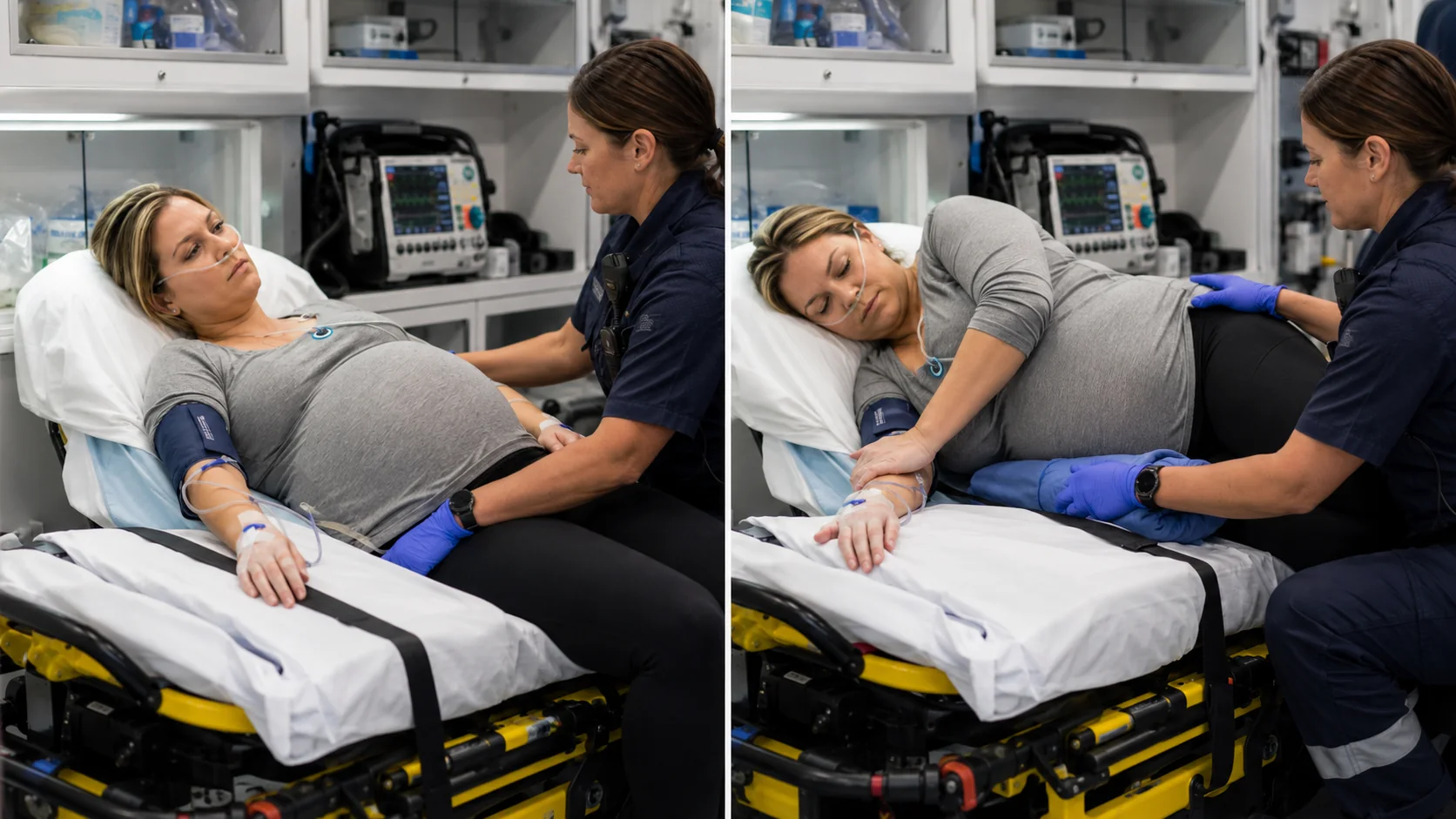
Mechanical compression remains
- Inferior vena cava compressed
- Venous return and cardiac output fall
- Uteroplacental perfusion may decline
- Maternal nausea, pallor, hypotension, or fetal deterioration may appear
Great-vessel pressure is relieved
- Venous return improves
- Cardiac output and pressure may recover
- Uterine perfusion improves
- Use manual left uterine displacement when the patient must remain supine
Transport actionRelieve the obstruction first, then reassess before escalating fluids or vasopressors.
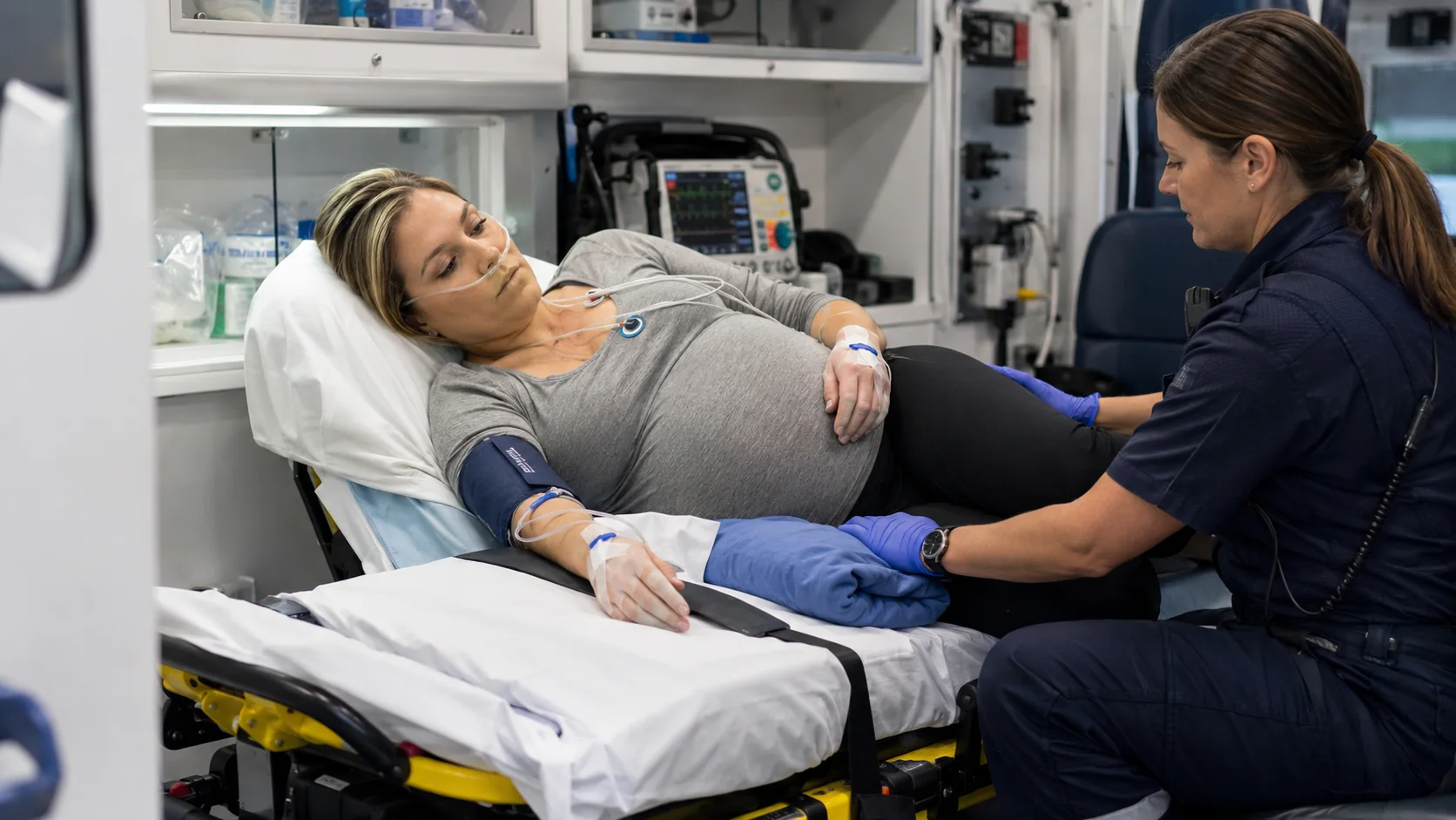
After approximately 20 weeks’ gestation, the enlarged uterus can compress the inferior vena cava when the patient lies supine. Venous return falls, reducing preload, stroke volume, cardiac output, and uterine perfusion. Some patients compensate through collateral circulation; others develop supine hypotensive syndrome with nausea, diaphoresis, pallor, dizziness, tachycardia, hypotension, altered mental status, or fetal heart-rate deterioration.
The aorta may also be compressed, particularly during profound hypotension or resuscitation, further reducing distal and uterine blood flow.
Transport intervention
- Place the patient in a left lateral tilt when clinically feasible.
- Use manual left uterine displacement when supine positioning is required, such as spinal motion restriction, airway management, or cardiopulmonary resuscitation.
- Reassess maternal blood pressure, symptoms, and fetal heart rate after repositioning.
- Secure the patient so the tilt is maintained during acceleration, braking, turns, and vibration.
2.5 Cardiac examination findings
Normal pregnancy can produce:
- A more forceful apical impulse
- A benign systolic flow murmur
- Mild peripheral edema
- Increased heart-rate variability with activity
Findings that require further evaluation include:
- A diastolic murmur
- Persistent resting hypoxemia
- Syncope with exertion
- Chest pain
- New pathologic dysrhythmia
- Pulmonary edema
- Marked jugular venous distention
- Unilateral leg swelling or pain
- Severe orthopnea or paroxysmal nocturnal dyspnea
Do not attribute all edema, tachycardia, or dyspnea to pregnancy.
3. Respiratory changes
Ventilation rises; oxygen reserve falls
Pregnancy changes the meaning of “normal”
Clinical interpretationPaCO₂ in the high 20s to low 30s mm Hg is expected. A value near 40 mm Hg may represent relative hypoventilation in a tiring patient.
Pregnancy increases oxygen demand while reducing oxygen reserve. Progesterone stimulates ventilation, the diaphragm elevates as the uterus enlarges, tidal volume rises, and functional residual capacity falls. Minute ventilation increases primarily through a larger tidal volume rather than a dramatic respiratory-rate increase.[1,5]
3.1 Increased minute ventilation and compensated respiratory alkalosis
A normal pregnant patient typically has a lower arterial carbon dioxide tension than a nonpregnant adult. PaCO₂ often falls into the high 20s to low 30s mm Hg, with renal bicarbonate loss producing metabolic compensation. The resulting pH is usually mildly alkalemic or high-normal.[1,5]
Paramedic interpretation
A PaCO₂ of 40 mm Hg may be normal in a nonpregnant adult but can indicate relative hypoventilation in a late-pregnancy patient who normally runs lower. In a critically ill pregnant patient, “normal” carbon dioxide may therefore be concerning if accompanied by fatigue, declining mental status, rising EtCO₂, or worsening work of breathing.
Capnography must be interpreted with ventilation, perfusion, and the clinical trajectory. Low EtCO₂ may reflect normal pregnancy hyperventilation, pain, anxiety, pulmonary embolism, sepsis, metabolic acidosis, or poor perfusion. A rising EtCO₂ in a tiring patient can signal failing ventilation even before frank hypercapnia is measured.
3.2 Reduced functional residual capacity
Functional residual capacity decreases as the diaphragm is displaced upward. At the same time, oxygen consumption rises. Together, these changes mean that apnea causes oxygen saturation to fall more quickly than in a comparable nonpregnant adult.[1,5]
Airway consequence
- Preoxygenation is essential.
- Apneic oxygenation should be considered when appropriate.
- Optimize position before induction.
- Prepare suction and backup airway devices before medications are given.
- Avoid repeated low-yield attempts.
- Confirm ventilation continuously with waveform capnography.
A patient who starts with a normal SpO₂ can desaturate rapidly after induction, seizure, opioid-related hypoventilation, or equipment disconnection.
3.3 Dyspnea in normal pregnancy
A sensation of breathlessness is common, especially with exertion. Normal pregnancy should not produce persistent hypoxemia, cyanosis, severe wheezing, focal crackles, hemoptysis, hypotension, marked tachypnea, or inability to speak in full sentences.
Red flags during transport
- SpO₂ below the expected normal range or a downward trend
- New oxygen requirement
- Respiratory rate persistently rising
- Increasing EtCO₂ after prior tachypnea
- Unilateral breath sounds
- Pulmonary edema or frothy sputum
- Pleuritic pain, syncope, or signs of venous thromboembolism
- Severe hypertension with dyspnea
- Fever and suspected infection
4. Airway and gastrointestinal changes
The pregnant airway has less margin for error
Prepare for difficulty before induction
Airway edema, reduced oxygen reserve, and aspiration risk can occur together. First-attempt success matters.
Airway edema
Smaller glottic opening, friable mucosa, and more difficult visualization.
Plan a smaller endotracheal tube.Rapid desaturation
Higher oxygen consumption plus lower functional residual capacity shortens safe apnea time.
Maximize preoxygenation.Aspiration risk
Reduced lower-esophageal sphincter tone and increased abdominal pressure promote regurgitation.
Test suction before medications.First-attempt planOptimize position · use the most experienced operator · video laryngoscopy when available · second-generation supraglottic airway ready · waveform capnography · failed-oxygenation plan.
4.1 Airway edema and difficult laryngoscopy
Pregnancy increases vascularity and edema of the upper airway. Nasal passages may be congested, mucosa can bleed easily, and the glottic opening may be smaller. Airway edema can worsen with preeclampsia, prolonged labor, aggressive fluid administration, or repeated airway manipulation.[1,6]
Transport implications
- Anticipate a potentially more difficult airway.
- Consider a smaller endotracheal tube than usual.
- Avoid traumatic nasal instrumentation when alternatives exist.
- Have a second-generation supraglottic airway available.
- Use the most experienced airway operator for the first attempt when possible.
- Prepare for a rapid transition from “difficult intubation” to “failed oxygenation.”
4.2 Aspiration risk
Reduced lower-esophageal sphincter tone, increased intra-abdominal pressure, labor, pain, opioids, and delayed gastric emptying in some circumstances increase regurgitation and aspiration risk.[1,6]
Practical precautions
- Position appropriately.
- Keep suction immediately available and tested.
- Avoid unnecessary oral intake during unstable transport.
- Use a deliberate rapid-sequence strategy when indicated.
- Recognize that aspiration can rapidly worsen maternal oxygenation and therefore fetal oxygen delivery.
5. Hematologic and coagulation changes
Pregnancy-specific context
System adaptations change how “normal” is interpreted
Use trends and the pregnancy baseline when interpreting laboratory values, fluid tolerance, metabolic emergencies, and medication clearance.
Hematologic
- Plasma expansion exceeds red-cell expansion
- Physiologic dilutional anemia
- Hypercoagulability and increased VTE risk
Pearl: fetal deterioration may precede maternal hypotension.
Renal and fluid
- Renal blood flow and GFR increase
- Expected creatinine is lower
- Lower oncotic pressure increases pulmonary-edema risk
Pearl: oliguria increases magnesium-toxicity risk.
Gastrointestinal
- Lower-esophageal sphincter tone decreases
- Intra-abdominal pressure rises
- Labor, opioids, and illness increase aspiration risk
Pearl: tested suction is part of airway preparation.
Metabolic and medications
- Metabolic demand and insulin resistance increase
- DKA may occur at lower glucose levels
- Volume of distribution and renal clearance change
Pearl: verify pump settings and rescue medications.
5.1 Physiologic anemia
Plasma volume expansion exceeds the increase in red-cell mass. The resulting dilution lowers measured hemoglobin and hematocrit compared with the nonpregnant baseline.[1,4]
This is not the same as iron-deficiency anemia, acute blood loss, or hemolysis. Interpretation should include gestational age, prior values, mean corpuscular volume, symptoms, bleeding, and the clinical trajectory.
5.2 Hypercoagulability
Pregnancy is a hypercoagulable state. Procoagulant factors increase, natural anticoagulant activity changes, and venous stasis develops—particularly in the lower extremities. The risk of venous thromboembolism is increased during pregnancy and remains elevated postpartum.[1,4]
Why this matters to paramedics
Pulmonary embolism must remain in the differential for unexplained dyspnea, pleuritic pain, syncope, tachycardia, hypoxemia, or hypotension. Do not dismiss these signs as normal pregnancy physiology.
5.3 Fibrinogen and hemorrhage
Fibrinogen normally rises during pregnancy. Therefore, a fibrinogen level that appears “normal” by nonpregnant standards may be relatively low in major obstetric hemorrhage or disseminated intravascular coagulation. Trend interpretation and obstetric context matter.
In the transport environment, you may not have real-time coagulation results. Clinical clues include ongoing bleeding, oozing from access sites, bruising, shock, placental abruption, retained dead fetus, amniotic fluid embolism, severe preeclampsia, HELLP syndrome, or massive transfusion.
6. Renal and fluid changes
Renal blood flow and glomerular filtration increase during normal pregnancy. Serum creatinine is therefore usually lower than in a nonpregnant adult.[7]
6.1 Creatinine interpretation
A creatinine value that looks “normal” on a general laboratory reference range may represent renal dysfunction in pregnancy. Always compare with prior values and the obstetric context.
6.2 Urine output
Urine output is an important transport trend, especially during:
- Magnesium sulfate infusion
- Preeclampsia or HELLP syndrome
- Hemorrhage
- Sepsis
- Diabetic ketoacidosis
- Renal disease
- Major trauma
Oliguria may indicate reduced renal perfusion, renal injury, severe preeclampsia, dehydration, or medication accumulation risk. In a patient receiving magnesium, declining urine output increases the risk of toxicity because magnesium is renally cleared.
6.3 Fluid balance and pulmonary edema
Pregnancy lowers colloid oncotic pressure and can increase susceptibility to pulmonary edema when hydrostatic pressure rises or capillary permeability is abnormal. Preeclampsia, cardiac disease, renal dysfunction, sepsis, tocolytics, and excessive crystalloid administration add risk.[1–3]
Transport principle
Treat the underlying physiology rather than reflexively administering large fluid volumes. A hypotensive patient may need blood products, hemorrhage control, relief of vena-caval compression, vasopressor support, or correction of sepsis—not simply repeated crystalloid boluses.
7. Endocrine, metabolic, and temperature considerations
Pregnancy creates increased metabolic demand and altered glucose handling. Insulin resistance rises later in pregnancy, while fasting glucose may be lower because of continuous maternal-fetal glucose use. Severe illness can destabilize glucose rapidly.
7.1 Diabetic ketoacidosis
Pregnant patients can develop diabetic ketoacidosis at lower glucose concentrations than expected. Vomiting, infection, insulin interruption, corticosteroids, beta-agonists, and dehydration may contribute. Do not exclude ketoacidosis solely because the glucose is not extremely elevated.
7.2 Temperature and fetal effects
Maternal fever increases maternal metabolic demand and can produce fetal tachycardia. Hypothermia can worsen coagulopathy in hemorrhage. Maintain normothermia during transport, especially in trauma, hemorrhage, sepsis, and delivery.
8. Musculoskeletal and anatomic changes relevant to transport
The enlarging uterus shifts the center of gravity and changes spinal curvature. Ligamentous laxity increases. These changes can affect safe lifting, stretcher positioning, spinal alignment, and patient comfort.
Practical implications
- Use adequate personnel for movement and loading.
- Avoid unsecured pillows or improvised wedges that can shift during transport.
- Reassess straps after positioning changes.
- Account for abdominal size when selecting restraint placement.
- Avoid direct compression over the uterus.
- In trauma, preserve spinal alignment while also relieving aortocaval compression.
9. Medication handling in pregnancy
Normal pregnancy can alter drug distribution and elimination through increased plasma volume, expanded total body water, lower albumin, altered hepatic enzyme activity, and increased renal filtration.[8]
These changes do not produce one universal dosing rule. Some medications may have a larger volume of distribution, altered free-drug fraction, or faster renal clearance. Clinical effect, protocol, infusion concentration, renal function, and maternal-fetal indication remain more important than memorizing a single pregnancy adjustment.
Transport medication safety
- Verify the exact concentration and pump settings at bedside before departure.
- Confirm whether the indication is maternal treatment, fetal benefit, or both.
- Know the expected maternal vital-sign effect.
- Know what fetal or neonatal effect should be anticipated.
- Identify the rescue medication before transport begins.
- Confirm how long the infusion can safely continue if diversion occurs.
- Reassess after every movement, pump transfer, power change, and tubing repositioning.
10. Normal findings versus dangerous findings
| Finding | May be physiologic | Concerning features |
|---|---|---|
| Heart rate | Mild increase from baseline | Persistent marked tachycardia, dysrhythmia, chest pain, syncope, shock signs |
| Blood pressure | Modest mid-pregnancy decrease | Severe hypertension, hypotension, narrow pulse pressure, worsening trend |
| Dyspnea | Mild exertional breathlessness | Hypoxemia, crackles, wheeze, hemoptysis, pleuritic pain, inability to speak normally |
| Edema | Mild bilateral dependent edema | Pulmonary edema, unilateral swelling, severe headache, hypertension, neurologic symptoms |
| Systolic murmur | Soft flow murmur | Diastolic murmur, loud pathologic murmur, cyanosis, heart-failure signs |
| Hemoglobin | Mild dilutional decrease | Rapid decline, bleeding, hemolysis, symptoms, hemodynamic change |
| Creatinine | Lower than nonpregnant baseline | Rising value or “normal-high” value in the wrong clinical context |
| PaCO₂ | High 20s to low 30s mm Hg | Rising toward nonpregnant normal with fatigue, acidosis, or respiratory distress |
11. Transport assessment checklist
Before departure and after every significant change, document or verify:
Maternal
- Position and response to position
- Mental status
- Heart rate and rhythm
- Blood pressure trend
- Respiratory rate and work of breathing
- SpO₂ and oxygen requirement
- EtCO₂ when indicated
- Lung sounds
- Skin findings and perfusion
- Pain and contractions
- Vaginal bleeding or fluid loss
- Uterine tenderness or tone
- IV/IO access and infusion status
- Urine output when monitored
Fetal
- Gestational age and viability context
- Baseline fetal heart rate
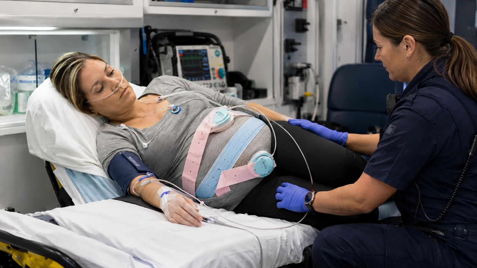
- Variability when continuous monitoring is available
- Accelerations or decelerations
- Relationship to contractions
- Change after maternal repositioning, oxygenation, or hemodynamic correction
Operational
- Remaining travel time
- Nearest appropriate alternate facility
- Obstetric and neonatal capability at each option
- Blood-product availability
- Delivery readiness
- Airway and suction readiness
- Communication plan if maternal or fetal status worsens
12. Evolving case study
Phase 1: Initial presentation
The 33-week patient from the opening scenario becomes hypotensive after being placed supine. You move the uterus leftward and elevate the left side of the stretcher. Her blood pressure improves to 104/64 mm Hg, heart rate decreases to 102/min, nausea resolves, and fetal heart rate returns to 140/min.
Interpretation: The rapid response supports aortocaval compression as the main mechanism. Continue to evaluate for additional causes because positional hypotension can coexist with hemorrhage, sepsis, preeclampsia, or medication effects.
Phase 2: New respiratory change
Thirty minutes later, she reports worsening dyspnea. Respiratory rate is 28/min, SpO₂ is 93%, and bilateral crackles are present. Blood pressure is 168/110 mm Hg. She has received 2 liters of crystalloid at the sending facility.
Interpretation: This is not normal pregnancy breathlessness. Severe hypertension, new hypoxemia, crackles, and substantial prior fluid raise concern for pulmonary edema associated with severe preeclampsia or cardiac dysfunction.
Phase 3: Transport decision
The original destination is 35 minutes away. A hospital with obstetric capability, anesthesia, blood bank, and emergency cesarean capability is 8 minutes away, though it lacks the originally requested subspecialty service.
Decision: Contact medical control and the receiving systems immediately. Maternal respiratory deterioration and potential fetal compromise may make diversion the safer option. The correct destination is determined by time, current instability, required immediate capability, and regional transfer agreements—not by the original plan alone.
13. High-yield chapter summary
- Pregnancy is a high-output, low-resistance physiologic state.
- Cardiac output and blood volume increase, while systemic vascular resistance falls.
- Plasma volume rises more than red-cell mass, producing physiologic anemia.
- Maternal hypotension can be late; fetal deterioration may occur first.
- Supine positioning after mid-pregnancy can reduce venous return and uteroplacental perfusion.
- Minute ventilation increases and normal PaCO₂ is lower than in a nonpregnant adult.
- Reduced functional residual capacity and increased oxygen consumption cause rapid desaturation during apnea.
- Upper-airway edema and aspiration risk make airway management more difficult.
- Pregnancy is hypercoagulable; pulmonary embolism must remain in the differential.
- Glomerular filtration increases, so serum creatinine is usually lower.
- Pulmonary edema risk rises with preeclampsia, cardiac disease, renal dysfunction, sepsis, and excessive fluid.
- Maternal positioning, oxygenation, ventilation, and perfusion are fetal interventions.
- Trends are more useful than isolated numbers.
- “Normal pregnancy” should never be used to explain hypoxemia, severe distress, syncope, pathologic murmurs, or progressive instability.
14. Embedded knowledge checks
A 30-week patient becomes nauseated and hypotensive after being secured flat on the stretcher. What should you do first?
Answer: Relieve aortocaval compression with left lateral tilt or manual left uterine displacement, then reassess maternal and fetal response. Continue evaluating for other causes if hypotension persists.
A late-pregnancy patient with respiratory distress has a PaCO₂ of 40 mm Hg. Why may this be concerning?
Answer: Normal pregnancy lowers PaCO₂ through increased minute ventilation. A value of 40 mm Hg may represent relative hypoventilation, particularly when paired with fatigue, rising EtCO₂, altered mentation, or worsening work of breathing.
Why can fetal heart-rate deterioration precede maternal hypotension in hemorrhage?
Answer: Maternal compensation may maintain systemic blood pressure while uterine perfusion falls. The placenta and fetus may therefore experience reduced oxygen delivery before obvious maternal cardiovascular collapse.
Why is a serum creatinine of 1.0 mg/dL potentially more concerning in pregnancy than in a nonpregnant adult?
Answer: Glomerular filtration normally increases and serum creatinine usually falls. A value that appears acceptable on a general laboratory range may represent a meaningful decline from the expected pregnancy baseline.
References
- Soma-Pillay P, Nelson-Piercy C, Tolppanen H, Mebazaa A. Physiological changes in pregnancy. Cardiovascular Journal of Africa. 2016;27(2):89–94. https://pmc.ncbi.nlm.nih.gov/articles/PMC4928162/
- Sanghavi M, Rutherford JD. Cardiovascular physiology of pregnancy. Circulation. 2014;130(12):1003–1008. https://doi.org/10.1161/CIRCULATIONAHA.114.009029
- Mehta LS, Warnes CA, Bradley E, et al. Cardiovascular considerations in caring for pregnant patients: a scientific statement from the American Heart Association. Circulation. 2020;141:e884–e903. https://doi.org/10.1161/CIR.0000000000000772
- Chandra M, et al. Natural physiological changes during pregnancy. 2024. https://pmc.ncbi.nlm.nih.gov/articles/PMC10964813/
- LoMauro A, Aliverti A. Respiratory physiology of pregnancy. Breathe. 2015;11(4):297–301. https://pmc.ncbi.nlm.nih.gov/articles/PMC4818213/
- Monanian G, et al. Anesthetic management of the pregnant patient undergoing non-obstetric surgery. 2025. https://pmc.ncbi.nlm.nih.gov/articles/PMC12028341/
- Cheung KL, Lafayette RA. Renal physiology of pregnancy. Advances in Chronic Kidney Disease. 2013;20(3):209–214. https://pmc.ncbi.nlm.nih.gov/articles/PMC4089195/
- Eke AC, et al. Physiologic changes during pregnancy and impact on drug disposition. 2023. https://pmc.ncbi.nlm.nih.gov/articles/PMC10365893/
- International Board of Specialty Certification. Maternal Fetal Transport Microcredential Candidate Handbook. Updated April 2026.
- Society for Maternal-Fetal Medicine. A maternal transport briefing form and checklist. Reaffirmed 2025. https://publications.smfm.org/publications/335-society-for-maternal-fetal-medicine-special-statement-a/
Chapter assessment
Twenty-question physiology quiz
Immediate rationales are shown in study mode, and your final score is stored locally.